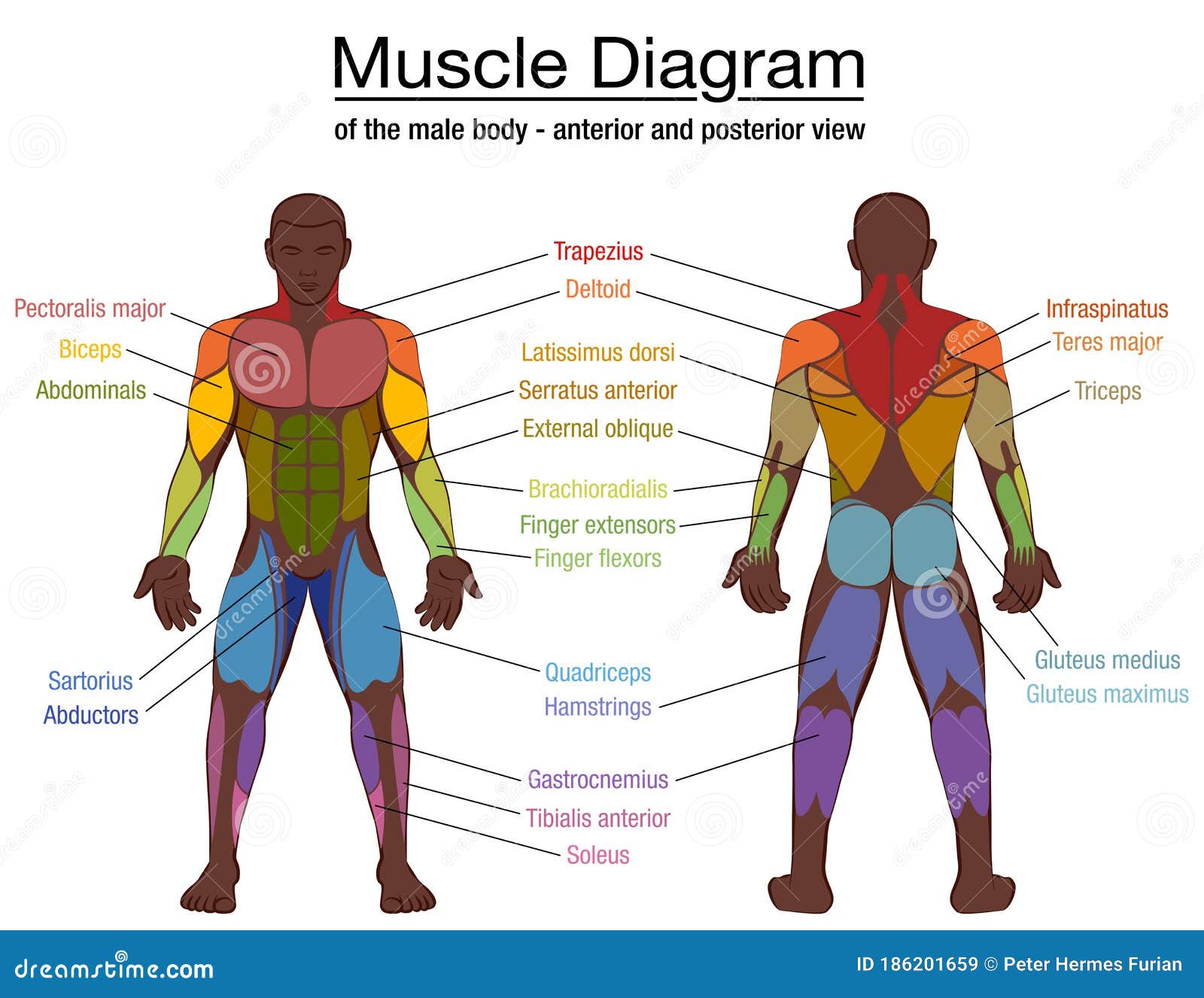
Chest Muscles Anatomy Labeled - Muscle Diagram Black Man Male Body Names Stock Vector Illustration Of Chest Packs 186201659 / (1) the pectoralis major, and (2) the pectoralis minor.
Chest Muscles Anatomy Labeled - Muscle Diagram Black Man Male Body Names Stock Vector Illustration Of Chest Packs 186201659 / (1) the pectoralis major, and (2) the pectoralis minor.. Ventral trunk muscles (overview) the trunk (torso) is the central part of the body to which the head and the limbs are attached. It forms the bony framework for breathing. It contains four muscles that exert a force on the upper limb: You must identify muscles based upon the names provided in the dissection study guide. Origin, insertion, innervation, and function should be labeled.
Learn about each of these muscles, their locations, functional anatomy and exercises for them. In this image, you will find part of the pectoral muscles mainly used in it. The pectoralis major originates along the clavicle, down the sternum, and across the ribs and inserts into the humerus. The muscles of the chest and upper back occupy the thoracic region of the body inferior to the neck and superior to the abdominal region and include the muscles of the shoulders. This is important to mention because it confuses many medical and physical therapy students because muscle group assignments not play a decisive role while studying.
Cat Chest Muscles Labeled from anatomycorner.com The pectoralis major, pectoralis minor, serratus anterior and subclavius. Ventral trunk muscles (overview) the trunk (torso) is the central part of the body to which the head and the limbs are attached. Compresses abdomen and rotates vertebral column. Chest muscles anatomy the chest is made up primarily of two muscles: Each type of muscle tissue in the human. Jetzt neu oder gebraucht kaufen. The muscles of the chest and upper back occupy the thoracic region of the body inferior to the neck and superior to the abdominal region and include the muscles of the shoulders. Labeled anatomy chart of male biceps and chest muscle on.
Über 7 millionen englischsprachige bücher.
The pectoral region is located on the anterior chest wall. The pectoralis major is labeled pectoralis transversus, and the pectoantebrachialis is labeled pectoralis. The pectoralis major originates along the clavicle, down the sternum, and across the ribs and inserts into the humerus. The secondary chest wall muscles correspond with the ventral shoulder and chest muscles. Here is the same image with the chest muscles labeled. You must identify muscles based upon the names provided in the dissection study guide. There is a printable worksheet available for download here so you can take the quiz with pen and paper. Male shoulder and chest muscles labeled chart on white labeled human anatomy diagram of male shoulder, biceps, arm, and chest muscles frontal anterior view on a white background. Related posts of chest muscles diagram anatomy muscle arm. Except for the brain, the trunk houses all the vital organs of the human body. A chest muscle that pulls the arm in towards the body. See more ideas about anatomy, anatomy and physiology, muscle anatomy. Human anatomy diagram shoulder anatomy shoulder muscles shoulder muscles and chest.
Add to favorites 0 favs. Compresses abdomen and rotates vertebral column. Chest muscle anatomy labeled muscle … multiple labeled and unlabeled sections from gross anatomy, showing the major structures of the chest, heart, and mediastinum. In spite of its resistance, the cage is dynamic, allowing pulmonary ventilation to. Computed tomography (ct) of the chest can detect pathology that may not show up on a conventional chest radiograph(1).
Muscle Diagram Black Man Male Body Names Stock Vector Illustration Of Chest Packs 186201659 from thumbs.dreamstime.com (1) the pectoralis major, and (2) the pectoralis minor. The torso muscles attach to the skeletal core of the trunk, and depending on their location are divided into two large groups: Male shoulder and chest muscles labeled chart on white labeled human anatomy diagram of male shoulder, biceps, arm, and chest muscles frontal anterior view on a white background. Use the mouse scroll wheel to move the images up and down alternatively use the tiny arrows (>>) on both side of the image to move the images.>>) on both side of the image to move the images. The pectoralis major, pectoralis minor, serratus anterior and subclavius. Each of these muscles has its origin on the scapula and inserts around the head of the humerus. The dominant muscle in the upper chest is the pectoralis major. It forms the bony framework for breathing.
The chest anatomy includes the pectoralis major, pectoralis minor and the serratus anterior.
The thoracic cage is a component of the thoracic wall and encloses the majority of the structures of the respiratory system. The dominant muscle in the upper chest is the pectoralis major. Chest muscles anatomy labeled : Here, we break down the anatomy of your chest muscles. Supraspinatus, infraspinatus, subscapularis, and teres minor. There is a printable worksheet available for download here so you can take the quiz with pen and paper. Computed tomography (ct) of the chest can detect pathology that may not show up on a conventional chest radiograph(1). Chest muscles anatomy the chest is made up primarily of two muscles: Assoc prof craig hacking et al. Cat anatomy dissection guide superficial muscles. See more ideas about anatomy, anatomy and physiology, muscle anatomy. Muscle anatomy books free download 12 photos of the muscle anatomy books free download muscle anatomy books free download, human muscles, muscle anatomy books free download. This is an online quiz called muscles of the anterior chest.
Here is the same image with the chest muscles labeled. Tr a p e zius. Related posts of chest muscles diagram anatomy muscle arm. Use the mouse scroll wheel to move the images up and down alternatively use the tiny arrows (>>) on both side of the image to move the images.>>) on both side of the image to move the images. The dominant muscle in the upper chest is the pectoralis major.
Chest Anatomy What Are The Muscles And What Do They Do Openfit from cdn.prod.openfit.com It contains four muscles that exert a force on the upper limb: This mri chest (thorax) axial cross sectional anatomy tool is absolutely free to use. The chest or thorax is the region between the neck and diaphragm that encloses organs, such as the heart, lungs, esophagus, trachea, and thoracic diaphragm. Muscles the dominant muscle in the upper chest is the pectoralis major. Related posts of chest muscles diagram anatomy muscle arm. Here is the same image with the chest muscles labeled. The pectoralis major is labeled pectoralis transversus, and the pectoantebrachialis is labeled pectoralis. This article lists a series of labeled imaging anatomy cases by system and modality.
(1) the pectoralis major, and (2) the pectoralis minor.
This is one of the internal rotator muscles that attach the humerus and internally rotate the arm. The pectoralis major is labeled pectoralis transversus, and the pectoantebrachialis is labeled pectoralis. These important muscles control many motions that involve moving the arms and head — such as throwing a ball, looking up at the sky, and raising your hand. Supraspinatus, infraspinatus, subscapularis, and teres minor. You must identify muscles based upon the names provided in the dissection study guide. Compresses abdomen and rotates vertebral column. It forms the bony framework for breathing. Here is the same image with the chest muscles labeled. (1) the pectoralis major, and (2) the pectoralis minor. Chest muscle anatomy labeled muscle … multiple labeled and unlabeled sections from gross anatomy, showing the major structures of the chest, heart, and mediastinum. Muscle anatomy books free download 12 photos of the muscle anatomy books free download muscle anatomy books free download, human muscles, muscle anatomy books free download. Ventral trunk muscles (overview) the trunk (torso) is the central part of the body to which the head and the limbs are attached. Tr a p e zius.
The chest anatomy includes the pectoralis major, pectoralis minor and the serratus anterior chest muscles anatomy. Anatomy ct axial chest form no 1.


0 Komentar